Eye Examination
Eye care services
At Darling St Optometrix, our eye examinations are not limited to a spectacle prescription check.
We believe in a wholistic approach to vision and our Optometrist has extensive experience in: Binocular Vision and Children’s eye assessments, myopia control treatments, diagnosis, and management of all eye conditions, specialised and advanced contact lens fittings, emergency red eye treatment and co-management of eye surgery.
Eye Examination
The standard scheduled eye examination is set for 1 to 3 years. However, this will be customised for every patient based on their testing results.
A comprehensive eye examination usually takes 30-45minutes.
This will include: an overall eye and medical history, baseline entry examinations, vision and prescription check, binocular vision function, front eye health assessment, back eye health assessment, eye pressure check, retinal imaging and computerised visual field assessment with further imaging if required.
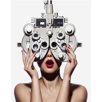
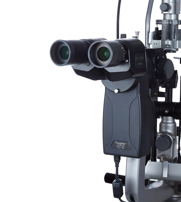
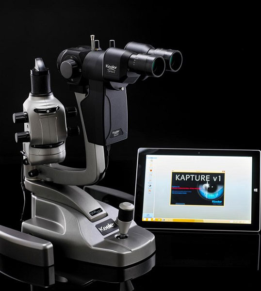
Slit Lamp Eye Assessment
A slit lamp is a microscope used to assess the health of the front of the eye. It helps us look at:
- Structure of the eyelids for any lesions, infections, or allergic reactions
- The state of the eyelashes and oil glands and tear ducts for any infections
- Conjunctiva and cornea for any inflammations, infections of foreign bodies
- Iris for any lesions
- Crystalline lens for cataracts
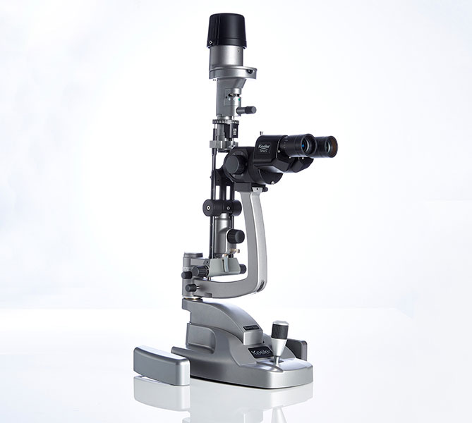
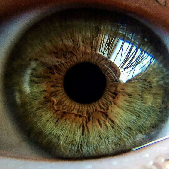
Anterior Slit Lamp Photography
Anterior eye photography, also known as anterior slit lamp photography, is a technique used to capture high-resolution images of the eye’s front structures, such as the cornea, iris, and lens.
Utilizing a slit lamp, this method allows eye care professionals to document and monitor conditions like cataracts, corneal abrasions, or inflammation. It is an essential tool for accurate diagnosis and ongoing management of eye health.
Digital Retinal Imaging
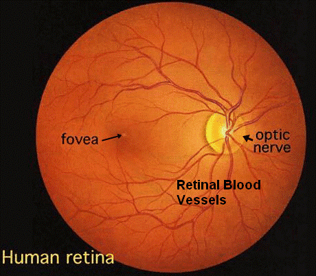
The Retina Scan Duo™ 2 includes a built-in 12-megapixel CCD camera, producing high-quality fundus images with a 45° angle of view. This advanced imaging captures detailed retinal structures, aiding in the early detection and management of conditions like macular degeneration and glaucoma.
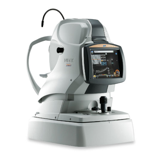
Ocular Coherence Tomography (OCT)
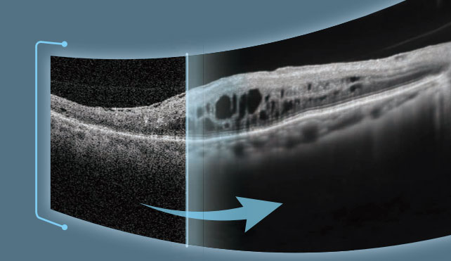
OCT is a 3-dimensional non-invasive imaging test which uses light waves to take cross-sectional (layered) scans of the retina. It is a It is used to provide additional information in the diagnosis of Glaucoma, Macula Degeneration, Diabetic Retinopathy and Macula swelling.
AngioScan
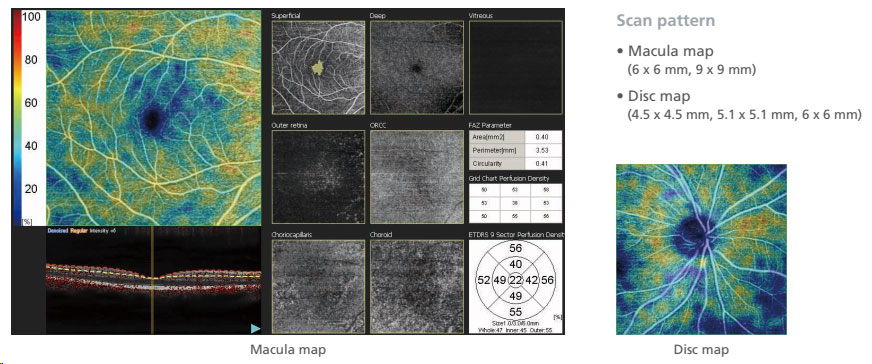
The optional AngioScan is available for OCT-Angiography imaging and diagnostics. The easy to use interface provides seven slabs for the macula map and four slabs for the disc map. This interface has intuitive functionality and removes projection artifacts. Segmentation into multiple slabs allows enhanced assessment of retinal microvasculature at specific depths and regions of interest. The effect of pathology can be evaluated in greater detail at each retinal depth.
Darling St Optometrix
At Darling St Optometrix, our eye examinations are not limited to a spectacle prescription check.
We believe in a wholistic approach to vision and our Optometrist has extensive experience in: Binocular Vision and Children’s eye assessments, myopia control treatments, diagnosis, and management of all eye conditions, specialised and advanced contact lens fittings, emergency red eye treatment and co-management of eye surgery.
Fundus autofluorescence (FAF)
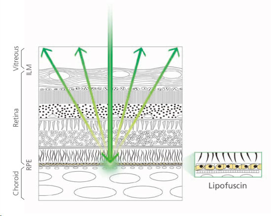
The FAF function is an advanced screening feature that allows non-invasive evaluation of the RPE without contrast dye.
FAF is naturally emitted due to the presence of a substance called lipofuscin in the RPE cells. When stimulated with a specific wavelength of light, lipofuscin fluoresces and its distribution can be mapped.
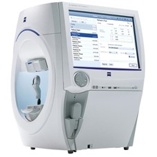
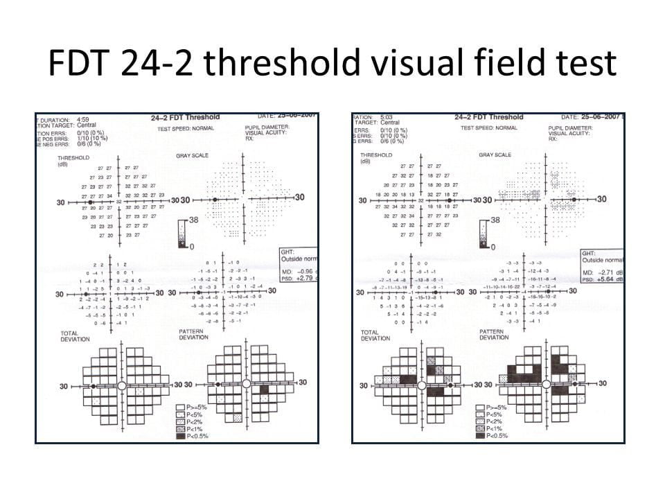
Computerised Visual Field Testing (VF)
Computerised Perimetry (Visual Field Testing) maps out the sensitivity of light at the back of the eye. This is useful in detecting both ocular and neurological condition.
Tonometry
Tonometry is a technique used to measure the pressure inside the eye.
The examination of eye pressure is an important test to assess inflammatory eye conditions, response to certain drugs and most importantly, the assessment of Glaucoma. However, alone, it is not a definitive diagnosis of Glaucoma.
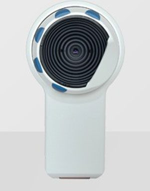
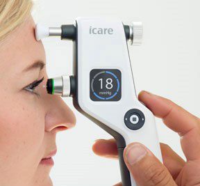
There are numerous instruments to measure eye pressure. Most patients are familiar with the ‘puff of air’ technique. We use a gentle, non-invasive instrument called i-care.
What is eye pressure?
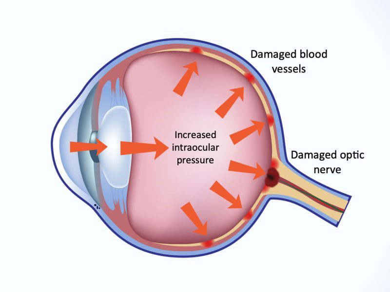
Towards the front of the eye, we have eye muscles which make fluid. The fluid circulates around the front of the eye and passes through a small drainage system. If this drainage system is narrow or blocked, there will be increased pressure forcing the fluid through. If the pressure rises, there is a stress effect towards the back of the eye around the exit hole. The nerves around the exit hole become compressed and lose function giving rise to reduced peripheral vision.
Normal eye pressure ranges between 15-21mmHg. It is measured using a form of pressure to indent the front of the eye and measure how long it takes to retract.
If the pressure is inside the eye is high, the retraction would be quick yielding a high reading.
If the pressure inside the eye is low, the retraction would be slow yielding a low reading.
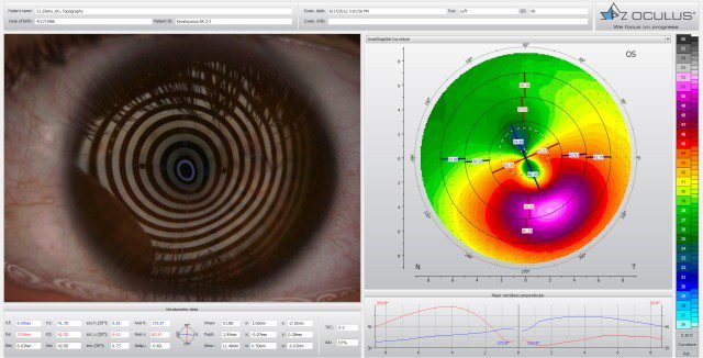
Corneal Topography
Corneal topography is a special 3D photography technique of the cornea (Front surface of the eye). It provides a detailed mapping of the cornea illustrating peaks and troughs, distortions and dryness of the cornea. All which create blurred vision unable to be corrected with spectacles.
Corneal topography is also used in special contact lens fittings such as Keratoconus, corneal graft lenses and Ortho-Keratology.
Corneal Pachymetry
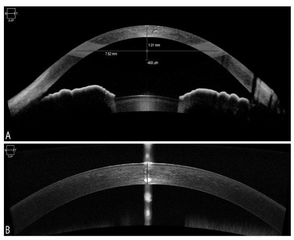
Corneal pachymetry is the measurement of the thickness of the cornea (front surface of the eye). This information is useful when assessing suitability for refractive(“laser”) surgery. It also provides important information when calibrating eye pressure in the diagnosis of Glaucoma.
