Keratoconus

What is Keratoconus?

Keratoconus is a common eye condition that affects the cornea – the front window of the eye. The cornea helps to bend and focus light to help us see.

In keratoconus, the cornea begins to thin. As a result, the round shape of the cornea gets distorted, and a cone-like shape develops. The reason for the cornea thinning and coning remains

When the cornea loses its shape, light can’t come to a sharp focus on the retina. This can cause blurry vision, double vision, astigmatism, short sightedness, or light sensitivity. If left untreated, vision will continue to get worse.

In most cases, keratoconus appears between the ages of 16 and 30 years and stabilises by middle age. Due to its early onset, it can impact school, work, social and family life if it’s not treated.

What causes Keratoconus?

The exact cause of keratoconus is not known. But both genetic and environmental factors seem to play a role.

Many people who are affected have no family history of the condition. But it is likely to have a genetic component.

Keratoconus often occurs alongside diseases where people have itchy eyes. For example, hay fever, allergies, and asthma. Lots of vigorous eye rubbing (Knuckle rubbing) can break down the fibres of the cornea. This might increase risk of keratoconus, but the exact link is still unclear.

How is Keratoconus diagnosed?

Patients usually present with blurred vision and sensitivity to light.

A routine eye examination will usually detect astigmatism and reduced vision.

Further investigation using corneal topography and pachymetry will diagnose Keratoconus.

How is Keratoconus treated by an Optometrist?

Initially Keratoconus is treated with spectacles and contact lenses.

Education on avoiding knuckle rubbing of the eyes and using anti histamine eye drops is also crucial to help reduce progression of the condition.

Monitoring of Keratoconus is the key factor to avoiding uncontrolled progression.

If vision cannot be corrected well with spectacles or soft contact lenses, RGP (Rigid Gas Permeable = Hard lenses) can be designed.

If there is progression of Keratoconus, referral to an Ophthalmologist is required.
How is Keratoconus treated by an Ophthalmologist?

Collagen Cross Linking:

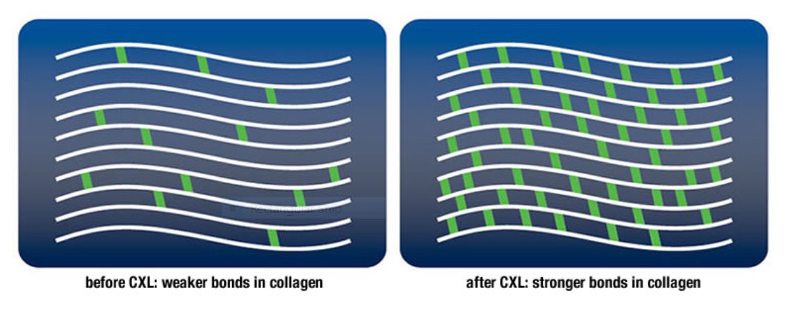
Corneal crosslinking increases the rigidity of a cornea that is weakened by keratoconus. The technique consists of applying a few drops of vitamin B2 (riboflavin) to the surface of the cornea so that the vitamin can penetrate. The cornea is subsequently exposed to a low ultraviolet light that causes a chemical reaction, which increases the thickness of the collagen fibres and the links between them, thus increasing the rigidity of the cornea.

It is currently the only procedure available that can slow or halt progressive keratoconus. Crosslinking may result in a slight flattening of the cornea, leading to small improvements in vision in certain cases. However, patients should not expect a reversal in existing vision loss from crosslinking nor should it be considered a cure for keratoconus.

Most patients in Australia with keratoconus are being treated today with crosslinking after being monitored for progression for a period of 3 to 6 months.

Intracorneal Ring Segments:

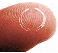
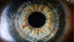
Intracorneal Ring Segments or ICRS are small plastic rings that are inserted into the cornea to flatten the conical shape of the cornea. They do NOT holt or minimise progression of Keratoconus. They merely manipulate the shape of the cornea to provide improvement in vision with/without spectacles. They are often known under their proprietary names of Intacs or Kerarings or Ferrara rings.

This mode of treatment has been available for over 10 years. The shape and design of the rings have been modified many times to achieve best results.

Kerarings are best used for early to moderate cases of keratoconus and specifically in patients who are contact lens intolerant. In recent years, Ophthalmologists have performed ring insertion in combination with cross linking in attempt to stabilise the condition.

In instances where Kera rings do not perform well, they can be removed by the Ophthalmologist.

Corneal Graft:

If Keratoconus is not diagnosed early or if it progresses without treatment, a corneal graft can be performed by an Ophthalmologist to restore vision. The technology and techniques available have vastly improved the results of this procedure.